Related articles
 胰岛素elisa试剂盒在操作的时候需要注意哪些事项?
胰岛素elisa试剂盒是固相夹心法酶联免疫吸附实验,已知待测物质浓度的标准品、未知浓度的样品加入微孔酶标板内进行检测。先将待测物质和生物素标记的抗体同时温育。洗涤后,加入亲和素标记过的HRP。...
ATCC细胞库在细胞培养上主要使用这个方法
ATCC细胞库的原理在不加任何条件下直接冻存细胞时,细胞内和外环境中的水都会形成冰晶,能导致细胞内发生机械损伤、电解质升高、渗透压改变、脱水、PH改变、蛋白变性等,能引起细胞死亡。如向培养液加入保护剂,可使冰点降低。...
用这个方法处理酶联免疫试剂盒的标本,效果更好
酶联免疫试剂盒实验中每一个步骤都尤为重要,细节不容忽视,它是实验成功的关键与否;在这里我司为方便各位了解,更好的完成实验,对酶联免疫试剂盒的实验原理和标本处理做出以下分析,供大家参考。...
关于人elisa试剂盒的组成结构,这里有详细说明
人elisa试剂盒运用双抗体夹心ELISA法定量测定兔血清、血浆、组织匀浆、细胞裂解液、细胞培养上清液和其他生物液体中兔Ⅰ型胶原α2(COL1α2)含量。...
HPLC检测与同类产品相比有以下优点
HPLC检测也称为高效液相色谱检测,是在传统液相色谱法的基础上发展而来的一种新一代色谱测定技术,HPLC将液体作为流动相,采用高压输液系统,将具有不同极性的单一溶剂或不同比例的混合溶剂、缓冲液等流动相泵入装有固定相的色谱柱...
分享感受态细胞的两种制作方法
感受态细胞的理化方法诱导细胞,吸收周围环境中的DNA分子,使其处于最适摄取和容纳外来DNA的生理状态。目前感受态细胞的主要原理就是通过处理使细胞的通透性变大,直观的说,使得细胞膜表面出现一些孔洞,便于外源基因或载体进入感受态细胞。...
关于ATCC细胞库的基础资料,以下有详细解析
ATCC细胞库管理包括,初级、主和工作细胞库。在某些特殊情况下,也可采用细胞种子和主细胞库二级管理,但需要得到国务院药品监督部门的批准。...
分享酶联elisa试剂盒操作前的严格要求方法
酶联elisa试剂盒操作前的严格要求方法,按试剂盒说明书的要求准备实验中需用的试剂。ELISA中用的蒸馏 水或去离子水,包括用于洗涤的,应为新鲜的和高质量的。...
上海elisa试剂盒是如何进行操作的?
上海elisa试剂盒是一种敏感性高,特异性强,重复性好的实验诊断方法。由于其试剂稳定、易保存,操作简便,结果判断较客观等因素,已广泛应用在植物学检验的各领域中。...
关于elisa实验步骤,我们已经为您整理好了
elisa是酶联接免疫吸附剂测定的简称。它是继免疫荧光和放射免疫技术之后发展起来的一种免疫酶技术。在检测时,受检标本与固相载体表面的抗体反应。洗涤后加入酶标记的抗体,通过反应结合在固相载体上。加入酶反应的底物后,底物被酶催化成为有色产物,产物的量与标本中受检物质的量直接相关,可根据呈色的深浅进行定性或定量分析。...
胰岛素elisa试剂盒在操作的时候需要注意哪些事项?
胰岛素elisa试剂盒是固相夹心法酶联免疫吸附实验,已知待测物质浓度的标准品、未知浓度的样品加入微孔酶标板内进行检测。先将待测物质和生物素标记的抗体同时温育。洗涤后,加入亲和素标记过的HRP。...
ATCC细胞库在细胞培养上主要使用这个方法
ATCC细胞库的原理在不加任何条件下直接冻存细胞时,细胞内和外环境中的水都会形成冰晶,能导致细胞内发生机械损伤、电解质升高、渗透压改变、脱水、PH改变、蛋白变性等,能引起细胞死亡。如向培养液加入保护剂,可使冰点降低。...
用这个方法处理酶联免疫试剂盒的标本,效果更好
酶联免疫试剂盒实验中每一个步骤都尤为重要,细节不容忽视,它是实验成功的关键与否;在这里我司为方便各位了解,更好的完成实验,对酶联免疫试剂盒的实验原理和标本处理做出以下分析,供大家参考。...
关于人elisa试剂盒的组成结构,这里有详细说明
人elisa试剂盒运用双抗体夹心ELISA法定量测定兔血清、血浆、组织匀浆、细胞裂解液、细胞培养上清液和其他生物液体中兔Ⅰ型胶原α2(COL1α2)含量。...
HPLC检测与同类产品相比有以下优点
HPLC检测也称为高效液相色谱检测,是在传统液相色谱法的基础上发展而来的一种新一代色谱测定技术,HPLC将液体作为流动相,采用高压输液系统,将具有不同极性的单一溶剂或不同比例的混合溶剂、缓冲液等流动相泵入装有固定相的色谱柱...
分享感受态细胞的两种制作方法
感受态细胞的理化方法诱导细胞,吸收周围环境中的DNA分子,使其处于最适摄取和容纳外来DNA的生理状态。目前感受态细胞的主要原理就是通过处理使细胞的通透性变大,直观的说,使得细胞膜表面出现一些孔洞,便于外源基因或载体进入感受态细胞。...
关于ATCC细胞库的基础资料,以下有详细解析
ATCC细胞库管理包括,初级、主和工作细胞库。在某些特殊情况下,也可采用细胞种子和主细胞库二级管理,但需要得到国务院药品监督部门的批准。...
分享酶联elisa试剂盒操作前的严格要求方法
酶联elisa试剂盒操作前的严格要求方法,按试剂盒说明书的要求准备实验中需用的试剂。ELISA中用的蒸馏 水或去离子水,包括用于洗涤的,应为新鲜的和高质量的。...
上海elisa试剂盒是如何进行操作的?
上海elisa试剂盒是一种敏感性高,特异性强,重复性好的实验诊断方法。由于其试剂稳定、易保存,操作简便,结果判断较客观等因素,已广泛应用在植物学检验的各领域中。...
关于elisa实验步骤,我们已经为您整理好了
elisa是酶联接免疫吸附剂测定的简称。它是继免疫荧光和放射免疫技术之后发展起来的一种免疫酶技术。在检测时,受检标本与固相载体表面的抗体反应。洗涤后加入酶标记的抗体,通过反应结合在固相载体上。加入酶反应的底物后,底物被酶催化成为有色产物,产物的量与标本中受检物质的量直接相关,可根据呈色的深浅进行定性或定量分析。...
| 品牌 | 自营品牌 | 货号 | BFN60800688 |
|---|---|---|---|
| 规格 | T25培养瓶x1 1.5ml冻存管x2 | 供货周期 | 现货 |
| 主要用途 | 仅供科研 | 应用领域 | 医疗卫生,生物产业 |
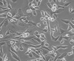
细胞名称 | 人肝癌细胞SK-HEP-1 |
| |
货物编码 | BFN60800688 | ||
产品规格 | T25培养瓶x1 | 1.5ml冻存管x2 | |
细胞数量 | 1x10^6 | 1x10^6 | |
保存温度 | 37℃ | -198℃ | |
运输方式 | 常温保温运输 | 干冰运输 | |
安全等级 | 1 | ||
用途限制 | 仅供科研用途 | ||
培养体系 | DMEM高糖培养基(Hyclone)+10%胎牛血清(Gibco)+1%双抗(Hyclone) | ||
培养温度 | 37℃ | 二氧化碳浓度 | 5% |
简介 | 人肝癌细胞SK-HEP-1细胞已被鉴定为内皮来源。该细胞系为异倍体女性人(XX),染色体在亚三倍体范围内。在裸鼠中,它能形成与肝癌相一致的大细胞癌。人肝癌细胞SK-HEP-1细胞由青旗(上海)生物技术发展有限公司于2017年引种自ATCC(HTB-52)。 | ||
注释 | Part of: Cancer Cell Line Encyclopedia (CCLE) project. Part of: COSMIC cell lines project. Part of: MD Anderson Cell Lines Project. From: Memorial Sloan Kettering Cancer Center; New York; USA. Registration: Memorial Sloan Kettering Cancer Center Office of Technology Development; SK1980-535. Characteristics: Has lost chromosome Y. Doubling time: 46.7 +- 10.3 hours, 94.2 hours (in CDM4-CHO medium), 289.8 hours (in 293 SFM II medium) (PubMed=25822314); ~30 hours (DSMZ). Microsatellite instability: Stable (MSS) (Sanger). Omics: Deep exome analysis. Omics: Deep RNAseq analysis. Omics: DNA methylation analysis. Omics: Protein expression by reverse-phase protein arrays. Omics: Secretome proteome analysis. Omics: SNP array analysis. Omics: Transcriptome analysis. | ||
STR信息 | Amelogenin:X,X;CSF1PO:11,12;D12S391:18,18;D13S317:8,12;D16S539:12,12;D18S51:13,15;D19S433:12,15.2;D21S11:29,31;D2S1338:20,23;D3S1358:16,16;D5S818:10,13;D6S1043:11,11;D7S820:8,11;D8S1179:13,14;FGA:17,17;PentaE:13,21;TH01:7,9;TPOX:9,9;vWA:14,17; | ||
参考文献 | DOI=10.1007/978-1-4757-1647-4_5 Fogh J., Trempe G.L. New human tumor cell lines. (In) Human tumor cells in vitro; Fogh J. (eds.); pp.115-159; Springer; New York (1975)
PubMed=327080; DOI=10.1093/jnci/59.1.221 Fogh J., Fogh J.M., Orfeo T. One hundred and twenty-seven cultured human tumor cell lines producing tumors in nude mice. J. Natl. Cancer Inst. 59:221-226(1977)
PubMed=833871; DOI=10.1093/jnci/58.2.209 Fogh J., Wright W.C., Loveless J.D. Absence of HeLa cell contamination in 169 cell lines derived from human tumors. J. Natl. Cancer Inst. 58:209-214(1977)
PubMed=924690; DOI=10.1002/ijc.2910200505 Kerbel R.S., Pross H.F., Leibovitz A. Analysis of established human carcinoma cell lines for lymphoreticular-associated membrane receptors. Int. J. Cancer 20:673-679(1977)
PubMed=6935474; DOI=10.1093/jnci/66.2.239 Wright W.C., Daniels W.P., Fogh J. Distinction of seventy-one cultured human tumor cell lines by polymorphic enzyme analysis. J. Natl. Cancer Inst. 66:239-247(1981)
PubMed=7017212; DOI=10.1093/jnci/66.6.1003 Pollack M.S., Heagney S.D., Livingston P.O., Fogh J. HLA-A, B, C and DR alloantigen expression on forty-six cultured human tumor cell lines. J. Natl. Cancer Inst. 66:1003-1012(1981)
PubMed=7459858 Rousset M., Zweibaum A., Fogh J. Presence of glycogen and growth-related variations in 58 cultured human tumor cell lines of various tissue origins. Cancer Res. 41:1165-1170(1981)
PubMed=6582512; DOI=10.1073/pnas.81.2.568 Mattes M.J., Cordon-Cardo C., Lewis J.L. Jr., Old L.J., Lloyd K.O. Cell surface antigens of human ovarian and endometrial carcinoma defined by mouse monoclonal antibodies. Proc. Natl. Acad. Sci. U.S.A. 81:568-572(1984)
PubMed=3518877; DOI=10.3109/07357908609038260 Fogh J. Human tumor lines for cancer research. Cancer Invest. 4:157-184(1986)
PubMed=2439335; DOI=10.1111/j.1432-1033.1987.tb11497.x Vincent C., Marceau M., Blangarin P., Bouic P., Madjar J.J., Revillard J.-P. Purification of alpha 1-microglobulin produced by human hepatoma cell lines. Biochemical characterization and comparison with alpha 1-microglobulin synthesized by human hepatocytes. Eur. J. Biochem. 165:699-704(1987)
PubMed=1371504; DOI=10.1007/BF02631017 Heffelfinger S.C., Hawkins H.H., Barrish J., Taylor L., Darlington G.J. SK HEP-1: a human cell line of endothelial origin. In Vitro Cell. Dev. Biol. Anim. 28:136-142(1992)
PubMed=8389256; DOI=10.1093/carcin/14.5.987 Hsu I.C., Tokiwa T., Bennett W., Metcalf R.A., Welsh J.A., Sun T., Harris C.C. p53 gene mutation and integrated hepatitis B viral DNA sequences in human liver cancer cell lines. Carcinogenesis 14:987-992(1993)
PubMed=9023415; DOI=10.1006/cimm.1996.1062 Seki N., Hoshino T., Kikuchi M., Hayashi A., Itoh K. HLA-A locus-restricted and tumor-specific CTLs in tumor-infiltrating lymphocytes of patients with non-small cell lung cancer. Cell. Immunol. 175:101-110(1997)
PubMed=9178645; DOI=10.1006/cimm.1997.1108 Nakao M., Sata M., Saitsu H., Yutani S., Kawamoto M., Kojiro M., Itoh K. CD4+ hepatic cancer-specific cytotoxic T lymphocytes in patients with hepatocellular carcinoma. Cell. Immunol. 177:176-181(1997)
PubMed=12029633; DOI=10.1053ep.2002.33683 Yasui K., Arii S., Zhao C., Imoto I., Ueda M., Nagai H., Emi M., Inazawa J. TFDP1, CUL4A, and CDC16 identified as targets for amplification at 13q34 in hepatocellular carcinomas. Hepatology 35:1476-1484(2002)
PubMed=12068308; DOI=10.1038/nature00766 Davies H., Bignell G.R., Cox C., Stephens P., Edkins S., Clegg S., Teague J.W., Woffendin H., Garnett M.J., Bottomley W., Davis N., Dicks E., Ewing R., Floyd Y., Gray K., Hall S., Hawes R., Hughes J., Kosmidou V., Menzies A., Mould C., Parker A., Stevens C., Watt S., Hooper S., Wilson R., Jayatilake H., Gusterson B.A., Cooper C., Shipley J.M., Hargrave D., Pritchard-Jones K., Maitland N.J., Chenevix-Trench G., Riggins G.J., Bigner D.D., Palmieri G., Cossu A., Flanagan A.M., Nicholson A., Ho J.W.C., Leung S.Y., Yuen S.T., Weber B.L., Seigler H.F., Darrow T.L., Paterson H., Marais R., Marshall C.J., Wooster R., Stratton M.R., Futreal P.A. Mutations of the BRAF gene in human cancer. Nature 417:949-954(2002)
PubMed=20069059; DOI=10.1155/2010/437143 Srisomsap C., Sawangareetrakul P., Subhasitanont P., Chokchaichamnankit D., Chiablaem K., Bhudhisawasdi V., Wongkham S., Svasti J. Proteomic studies of cholangiocarcinoma and hepatocellular carcinoma cell secretomes. J. Biomed. Biotechnol. 2010:437143-437143(2010)
PubMed=20164919; DOI=10.1038/nature08768 Bignell G.R., Greenman C.D., Davies H., Butler A.P., Edkins S., Andrews J.M., Buck G., Chen L., Beare D., Latimer C., Widaa S., Hinton J., Fahey C., Fu B., Swamy S., Dalgliesh G.L., Teh B.T., Deloukas P., Yang F., Campbell P.J., Futreal P.A., Stratton M.R. Signatures of mutation and selection in the cancer genome. Nature 463:893-898(2010)
PubMed=22460905; DOI=10.1038/nature11003 Barretina J.G., Caponigro G., Stransky N., Venkatesan K., Margolin A.A., Kim S., Wilson C.J., Lehar J., Kryukov G.V., Sonkin D., Reddy A., Liu M., Murray L., Berger M.F., Monahan J.E., Morais P., Meltzer J., Korejwa A., Jane-Valbuena J., Mapa F.A., Thibault J., Bric-Furlong E., Raman P., Shipway A., Engels I.H., Cheng J., Yu G.K., Yu J., Aspesi P. Jr., de Silva M., Jagtap K., Jones M.D., Wang L., Hatton C., Palescandolo E., Gupta S., Mahan S., Sougnez C., Onofrio R.C., Liefeld T., MacConaill L.E., Winckler W., Reich M., Li N., Mesirov J.P., Gabriel S.B., Getz G., Ardlie K., Chan V., Myer V.E., Weber B.L., Porter J., Warmuth M., Finan P., Harris J.L., Meyerson M., Golub T.R., Morrissey M.P., Sellers W.R., Schlegel R., Garraway L.A. The Cancer Cell Line Encyclopedia enables predictive modelling of anticancer drug sensitivity. Nature 483:603-607(2012)
PubMed=23505090; DOI=10.1002/hep.26402 Wang K., Lim H.Y., Shi S., Lee J., Deng S., Xie T., Zhu Z., Wang Y., Pocalyko D., Yang W.J., Rejto P.A., Mao M., Park C.-K., Xu J. Genomic landscape of copy number aberrations enables the identification of oncogenic drivers in hepatocellular carcinoma. Hepatology 58:706-717(2013)
PubMed=23887712; DOI=10.1038/ncomms3218 Nault J.-C., Mallet M., Pilati C., Calderaro J., Bioulac-Sage P., Laurent C., Laurent A., Cherqui D., Balabaud C., Zucman-Rossi J. High frequency of telomerase reverse-transcriptase promoter somatic mutations in hepatocellular carcinoma and preneoplastic lesions. Nat. Commun. 4:2218-2218(2013)
PubMed=25574106; DOI=10.3748/wjg.v21.i1.311 Cevik D., Yildiz G., Ozturk M. Common telomerase reverse transcriptase promoter mutations in hepatocellular carcinomas from different geographical locations. World J. Gastroenterol. 21:311-317(2015)
PubMed=25822314; DOI=10.1007/s00449-015-1392-9 Biaggio R.T., Abreu-Neto M.S., Covas D.T., Swiech K. Serum-free suspension culturing of human cells: adaptation, growth, and cryopreservation. Bioprocess Biosyst. Eng. 38:1495-1507(2015)
PubMed=27397505; DOI=10.1016/j.cell.2016.06.017 Iorio F., Knijnenburg T.A., Vis D.J., Bignell G.R., Menden M.P., Schubert M., Aben N., Goncalves E., Barthorpe S., Lightfoot H., Cokelaer T., Greninger P., van Dyk E., Chang H., de Silva H., Heyn H., Deng X., Egan R.K., Liu Q., Mironenko T., Mitropoulos X., Richardson L., Wang J., Zhang T., Moran S., Sayols S., Soleimani M., Tamborero D., Lopez-Bigas N., Ross-Macdonald P., Esteller M., Gray N.S., Haber D.A., Stratton M.R., Benes C.H., Wessels L.F.A., Saez-Rodriguez J., McDermott U., Garnett M.J. A landscape of pharmacogenomic interactions in cancer. Cell 166:740-754(2016)
PubMed=28196595; DOI=10.1016/j.ccell.2017.01.005 Li J., Zhao W., Akbani R., Liu W., Ju Z., Ling S., Vellano C.P., Roebuck P., Yu Q., Eterovic A.K., Byers L.A., Davies M.A., Deng W., Gopal Y.N.V., Chen G., von Euw E.M., Slamon D.J., Conklin D., Heymach J.V., Gazdar A.F., Minna J.D., Myers J.N., Lu Y., Mills G.B., Liang H. Characterization of human cancer cell lines by reverse-phase protein arrays. Cancer Cell 31:225-239(2017)
PubMed=30894373; DOI=10.1158/0008-5472.CAN-18-2747 Dutil J., Chen Z., Monteiro A.N., Teer J.K., Eschrich S.A. An interactive resource to probe genetic diversity and estimated ancestry in cancer cell lines. Cancer Res. 79:1263-1273(2019)
PubMed=31068700; DOI=10.1038/s41586-019-1186-3 Ghandi M., Huang F.W., Jane-Valbuena J., Kryukov G.V., Lo C.C., McDonald E.R. III, Barretina J., Gelfand E.T., Bielski C.M., Li H., Hu K., Andreev-Drakhlin A.Y., Kim J., Hess J.M., Haas B.J., Aguet F., Weir B.A., Rothberg M.V., Paolella B.R., Lawrence M.S., Akbani R., Lu Y., Tiv H.L., Gokhale P.C., de Weck A., Mansour A.A., Oh C., Shih J., Hadi K., Rosen Y., Bistline J., Venkatesan K., Reddy A., Sonkin D., Liu M., Lehar J., Korn J.M., Porter D.A., Jones M.D., Golji J., Caponigro G., Taylor J.E., Dunning C.M., Creech A.L., Warren A.C., McFarland J.M., Zamanighomi M., Kauffmann A., Stransky N., Imielinski M., Maruvka Y.E., Cherniack A.D., Tsherniak A., Vazquez F., Jaffe J.D., Lane A.A., Weinstock D.M., Johannessen C.M., Morrissey M.P., Stegmeier F., Schlegel R., Hahn W.C., Getz G., Mills G.B., Boehm J.S., Golub T.R., Garraway L.A., Sellers W.R. Next-generation characterization of the Cancer Cell Line Encyclopedia. Nature 569:503-508(2019) | ||
验收细胞注意事项
1、收到人肝癌细胞SK-HEP-1细胞,请查看瓶子是否有破裂,培养基是否漏出,是否浑浊,如有请尽快联系。
2、收到人肝癌细胞SK-HEP-1细胞,如包装完好,请在显微镜下观察细胞。,由于运输过程中的问题,细胞培养瓶中的贴壁细胞有可能从瓶壁中脱落下来,显微镜下观察会出现细胞悬浮的情况,出现此状态时,请不要打开细胞培养瓶,应立即将培养瓶置于细胞培养箱里静止 3-5 小时左右,让细胞先稳定下,再于显微镜下观察,此时多数细胞会重新贴附于瓶壁。如细胞仍不能贴壁,请用台盼蓝染色法鉴定细胞活力,如台盼蓝染色证实细胞活力正常请按悬浮细胞的方法处理。
3、收到人肝癌细胞SK-HEP-1细胞后,请镜下观察细胞,用恰当方式处理细胞。若悬浮的细胞较多,请离心收集细胞,接种到一个新的培养瓶中。弃掉原液,使用新鲜配制的培养基,使用进口胎牛血清。刚接到细胞,若细胞不多时 血清浓度可以加到 15%去培养。若细胞迏到 80%左右 ,血清浓度还是在 10%。
4、收到人肝癌细胞SK-HEP-1细胞时如无异常情况 ,请在显微镜下观察细胞密度,如为贴壁细胞,未超过80%汇合度时,将培养瓶中培养基吸出,留下 5-10ML 培养基继续培养:超过 80%汇合度时,请按细胞培养条件传代培养。如为悬浮细胞,吸出培养液,1000 转/分钟离心 3 分钟,吸出上清,管底细胞用新鲜培养基悬浮细胞后移回培养瓶。
5、将培养瓶置于 37℃培养箱中培养,盖子微微拧松。吸出的培养基可以保存在灭菌过的瓶子里,存放于 4℃冰箱,以备不时之需。
6、24 小时后,人肝癌细胞SK-HEP-1细胞形态已恢复并贴满瓶壁,即可传代。(贴壁细胞)将培养瓶里的培养基倒去,加 3-5ml(以能覆盖细胞生长面为准)PBS 或 Hanks’液洗涤后弃去。加 0.5-1ml 0.25%含 EDTA 的胰酶消化,消化时间以具体细胞为准,一般 1-3 分钟,不超过 5 分钟。可以放入37℃培养箱消化。轻轻晃动瓶壁,见细胞脱落下来,加入 3-5ml 培养基终止消化。用移液管轻轻吹打瓶壁上的细胞,使之*脱落,然后将溶液吸入离心管内离心,1000rpm/5min。弃上清,视细胞数量决定分瓶数,一般一传二,如细胞量多可一传三,有些细胞不易传得过稀,有些生长较快的细胞则可以多传几瓶,以具体细胞和经验为准。(悬浮细胞)用移液管轻轻吹打瓶壁,直接将溶液吸入离心管离心即可。
7、贴壁细胞 ,悬浮细胞。严格无菌操作。换液时,换新的细胞培养瓶和换新鲜的培养液,37℃,5%CO2 培养。
特别提醒: 原瓶中培养基不宜继续使用,请更换新鲜培养基培养。

 您的位置:
您的位置:

 咨询电话
咨询电话